

Translate this page into:
Diascopy Revisited: ‘Slide’ Your Way to Diagnosis
*Corresponding author: Priya Prafulla Kadu, Department of Dermatology, Venerology and Leprosy, Government Medical College, Akola, Maharashtra, India. kadupriya15@gmail.com
-
Received: ,
Accepted: ,
How to cite this article: Kadu PP. Diascopy Revisited: ‘Slide’ Your Way to Diagnosis. Indian J Postgrad Dermatol. 2024;2:17-9. doi: 10.25259/IJPGD_71_2023
INTRODUCTION
Andrews has defined the diascope as ‘a glass plate pressed against the skin for observing anatomic changes other than those of congestion.’[1] It is an important bedside diagnostic tool used daily to evaluate several dermatological conditions. Apart from skin, mucosae can also be examined using diascopy. It is a simple, rapid and non-invasive technique adjuvant in diagnosing dermatological disorders. It is also known as vitropression.[2]
TYPES OF DIASCOPE
There are three types of diascopes:[2]
The European curved type – made of glass
The plastic curved type – a modification of the European curved type
A glass slide
Modifications:
PROCEDURE
Firm pressure is applied with the help of a hard, transparent instrument such as a glass slide, plastic spatula [2] or two microscopic slides. The objects are pressed over the surface of the lesion to induce blanching. Due to the displacement of the blood from the vessel, a blanched appearance can be noted.[4,5] Glass ampoules can also be used.[2] The procedure is performed for 1–2 min, during which there will be emptying of the vessels, giving rise to a pale appearance. This indicates a positive test. The pressure required for the test is variable. Care should be taken to prevent injury to the skin, which may occur by breakage of the slide. To avoid this, two glass slides may be used. After removal of the slide, the lesion appears pale for a few seconds before there is a refilling of the blood from the vessel.
INDICATIONS
To differentiate a vascular lesion from a pigmented lesion
To differentiate purpura [Figure 1] (due to extravasation of RBCs) from erythema (due to dilatation of blood vessels)
To determine the central feeder vessel in telangiectasias[2]
To visualise the pigment spots over the post-operative site of vitiligo surgery[3]
For granulomatous disorders: In lupus vulgaris [Figure 2] to observe apple jelly nodules on blanching,[6] in sarcoidosis and tuberculosis to study the glassy brown appearance[7]
For differentiating urticaria (blanchable) from urticarial vasculitis (non-blanchable)[8]
To differentiate nevus anemicus from nevus depigmentosus and vitiligo. Nevus anemicus blanches on diascopy and the borders merge with surrounding normal skin whereas nevus depigmentosus and vitiligo remain the same[9]
To differentiate lip vitiligo having uniform convex margins from post-inflammatory hypopigmentation, which appears tan brown with uneven or jagged margins[2]
To make the lesions more conspicuous in lichen planus, lichen sclerosus et atrophicus, and seborrheic keratosis cases[8]
Useful in cases of xanthomas, necrobiosis lipoidica and pseudoxanthoma elasticum to make the yellow colour more noticeable[1]
A petri dish can be used to examine a pigmented lesion like a nevus or melanoma to enhance features. The procedure involves the application of liquid paraffin, water or any disinfectant to the skin lesion, followed by placement of the Petri dish over the lesion and illumination with a flashlight at 70° to the Petri dish.[10] A dermoscopy-like visualisation is possible using a magnifying lens[10]
Potentially identify vascular lesions of the oral cavity.[9]

- Use of diascopy to differentiate purpura from erythema.
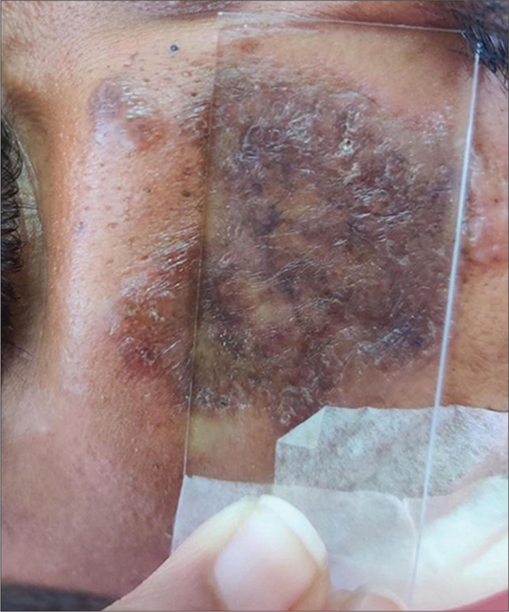
- Apple jelly nodules on diascopy in lupus vulgaris.
LIMITATIONS
Can only be used as an adjuvant in the diagnosis.
ADVANTAGES
Rapid, non-invasive and easy.
Ethical approval
The research was in compliance with Helsinki declaration 1964.
Declaration of patient consent
The authors certify that they have obtained all appropriate patient consent.
Conflicts of interest
There are no conflicts of interest.
Use of artificial intelligence (AI)-assisted technology for manuscript preparation
The author confirms that there was no use of artificial intelligence (AI)-assisted technology for assisting in the writing or editing of the manuscript and no images were manipulated using AI.
Financial support and sponsorship
Nil.
References
- Investigative and Clinical Studies with Diascopy in Dermatology. AMA Arch Derm. 1957;75:699-705.
- [CrossRef] [PubMed] [Google Scholar]
- Diascopy in Oral Lesions: An Old Algorithm Revisited. J Dent Oral Biol. 2017;2:1114.
- [Google Scholar]
- A Novel Technique of Diascopy Over Curved Surface of Body. Indian Dermatol Online J. 2023;14:297-8.
- [CrossRef] [PubMed] [Google Scholar]
- Diascopy: A Clinical Technique for the Diagnosis of Vascular Lesions. Gen Dent. 2001;49:206-9.
- [Google Scholar]
- Illustrated Synopsis of Dermatology and Sexually Transmitted Diseases (4th ed). Amsterdam: Elsevier; 2014.
- [Google Scholar]
- Glass Slide-An Indispensable Tool for the Dermatologist. CosmoDerma. 2022;2:27.
- [CrossRef] [Google Scholar]
- Clinical Value of Diascopy and Other Noninvasive Techniques on Differential Diagnosis Algorithms of Oral Pigmentations: A Systematic Review. J Clin Exp Dent. 2016;8:e448-58.
- [CrossRef] [PubMed] [Google Scholar]
- Diagnostic Pearl: Unmagnified Diascopy for Large Pigmented Lesions Reveals Features Similar to those of Epiluminescence Microscopy. J Am Acad Dermatol. 1999;41:765-6.
- [CrossRef] [PubMed] [Google Scholar]


