

Translate this page into:
Dermoscopy of Cutaneous Larva Migrans
*Corresponding author Arun Somasundaram, Department of Dermatology, Jawaharlal Institute of Postgraduate Medical Education and Research (JIPMER), Puducherry, India. arunsomasundaram25@gmail.com
-
Received: ,
Accepted: ,
How to cite this article: Somasundaram A, Murugan K. Dermoscopy of Cutaneous Larva Migrans. Indian J Postgrad Dermatol. 2024;2:52-3. doi: 10.25259/IJPGD_4_2024
A man in his 20s presented with itchy skin lesions over the right side of his trunk for 1-week duration. Cutaneous examination revealed multiple skin-coloured linear bizarre tracts and papules over the right side of the trunk extending to the arm and axilla [Figure 1a and b]. The rest of the mucocutaneous examination was non-contributory. A diagnosis of cutaneous larva migrans was made. The patient was initiated on tablet albendazole 400 mg along with tablet ivermectin for 3 days following which he had improvement in skin lesions [Figure 1c]. Dermoscopy examination revealed brownish linear serpiginous tracts suggestive of cutaneous larva migrans [Figure 2]. The brownish linear serpiginous tracts suggest a larval body and brownish dots represent an empty larval tract in the image [Figure 2]. Larva migrans is usually a diagnosis made clinically, however, dermoscopy can aid as an adjunct to the diagnosis.

- (a) Multiple skin-coloured linear bizarre tracts and papules over the right side of the trunk extending to the arm and axilla. (b) Close-up image showing serpiginous tracts of larva migrans. (c) Improvement in skin lesions noted on follow-up after 3 weeks.
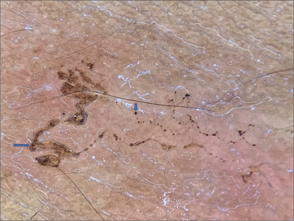
- Dermoscopy image (DermLite, × 10, polarised mode) revealed brownish linear serpiginous tracts (larval body) with brownish dots (empty larval tract) suggestive of cutaneous larva migrans.
Ethical approval
Institutional Review Board approval is not required.
Declaration of patient consent
The authors certify that they have obtained all appropriate patient consent.
Conflicts of interest
There are no conflicts of interest.
Use of artificial intelligence (AI)-assisted technology for manuscript preparation
The authors confirm that there was no use of artificial intelligence (AI)-assisted technology for assisting in the writing or editing of the manuscript and no images were manipulated using AI.
Financial support and sponsorship
Nil.


