

Translate this page into:
Flat Erythematous Lesion on Cheek
*Corresponding author: Niharika Dhattarwal, Department of Dermatology, Chacha Nehru Bal Chikitsalaya, New Delhi, India. niharika.pgirtk@gmail.com
-
Received: ,
Accepted: ,
How to cite this article: Dhattarwal N, Bansal S. Flat Erythematous Lesion on Cheek. Indian J Postgrad Dermatol. 2024;2:146-7. doi: 10.25259/IJPGD_111_2024
CASE DESCRIPTION
A 17-year-old female presented with reddish lesion on her left cheek [Figure 1]. The lesion was noted 6–7 years ago and is non-progressive and asymptomatic. There is no history of similar lesions elsewhere or any systemic complaints. On inspection, ill-defined erythematous lesion was seen on left cheek with no surface changes. On palpation, lesion was flat, non-pulsatile and local temperature was normal.
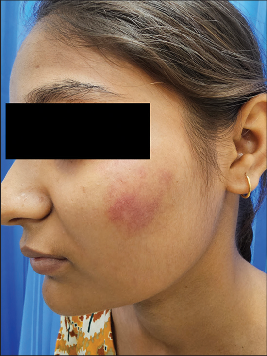
- Red coloured flat lesion present on the left cheek.
DERMOSCOPIC FINDINGS
On dermoscopy, multiple red tortuous capillaries are shown [Figure 2a]. On slightly pressing the dermoscope against the lesion, we noted that the lesions were blanchable [Figure 2b].

- (a) Multiple red tortuous capillaries seen on dermoscopy (DermLite DL4, non-polarised, × 40) and (b) blanching of lesion seen on ‘Dermoscopic diascopy’.
DIAGNOSIS
Unilateral nevoid telangiectasia.
DISCUSSION
Unilateral nevoid telangiectasia is a benign, vascular disorder having superficial, clustered telangiectasias in a unilateral linear fashion. Congenital form is predominant in males; acquired form is more commonly seen in women around puberty or during pregnancy and is believed to be triggered by an elevated level of or sensitivity to oestrogen. Clinically, numerous telangiectatic macules are seen along dermatomes or Blaschko’s lines. The lesions are blanchable on diascopy.[1] Histopathologically, dilated, thin-walled endothelium-lined vessels are seen in upper and mid dermis. There is no inflammation, neoangiogenesis or aberrant malformed vessels. Dermoscopic findings concur with the histology and normal dilated capillaries with a reticulated appearance are noted.
Differential diagnoses include angioma serpiginosum, circumscribed naviform angiokeratoma, hereditary benign telangiectasia, nevus vascularis mixtus and nevus flammeus. Angioma serpiginosum are capillary malformations presenting as punctate maculopapular lesions in a linear, serpiginous pattern. These do not blanch with diascopy and show presence of multiple red oval lagoons on dermoscopy.[2]
We have described ‘dermoscopic diascopy’ of this condition in our patient. Dermoscopic diascopy is a simple technique that effectively combines two bedside tests: Dermoscopy and diascopy can be utilised for various vascular and granulomatous disorders in dermatology.
Ethical approval
Institutional Review Board approval is not required.
Declaration of patient consent
The authors certify that they have obtained all appropriate patient consent.
Conflicts of interest
There are no conflicts of interest.
Use of artificial intelligence (AI)-assisted technology for manuscript preparation
The authors confirm that there was no use of artificial intelligence (AI)-assisted technology for assisting in the writing or editing of the manuscript and no images were manipulated using AI.
Financial support and sponsorship
Nil.
References
- Acquired Unilateral Nevoid Telangiectasia with Pruritus and Unknown Etiology. Cutis. 2021;107:E42-3.
- [CrossRef] [PubMed] [Google Scholar]
- Dermoscopy as an Important Tool for Differentiating Unilateral Nevoid Telangiectasia and Angioma Serpiginosum. Dermatol Pract Concept. 2019;9:306-7.
- [CrossRef] [PubMed] [Google Scholar]


