

Translate this page into:
Diascopy and Dermatoscopy of Lupus Vulgaris: The Tale of ‘Apple-jelly’ Nodules

*Corresponding author: Prakhar Srivastava, Department of Dermatology and STD, Vardhman Mahavir Medical College and Safdarjung Hospital, New Delhi, India. sriprakhar1996@gmail.com
-
Received: ,
Accepted: ,
How to cite this article: Srivastava P, Srivastava P, Khunger N. Diascopy and Dermatoscopy of Lupus Vulgaris: The Tale of ‘Apple-jelly’ Nodules. Indian J Postgrad Dermatol. 2024;2:54-6. doi:10.25259/IJPGD_88_2023
CASE DESCRIPTION AND DERMATOSCOPIC FINDINGS
A lady in her 40s presented with a reddish lesion on the right side of her face of 6 months duration. On examination, solitary, succulent, and reddish-brown plaque measuring 10 × 6 cm was present over the lower right cheek, with scarring at the inferior margin and serpiginous progression at the superior margin [Figure 1]. Diascopy showed apple-jelly nodules, and dermatoscopy revealed yellow-brown areas with overlying branching vessels and white scales [Figures 2 and 3]. Mantoux test was strongly positive. Histopathology showed epithelioid granulomas.
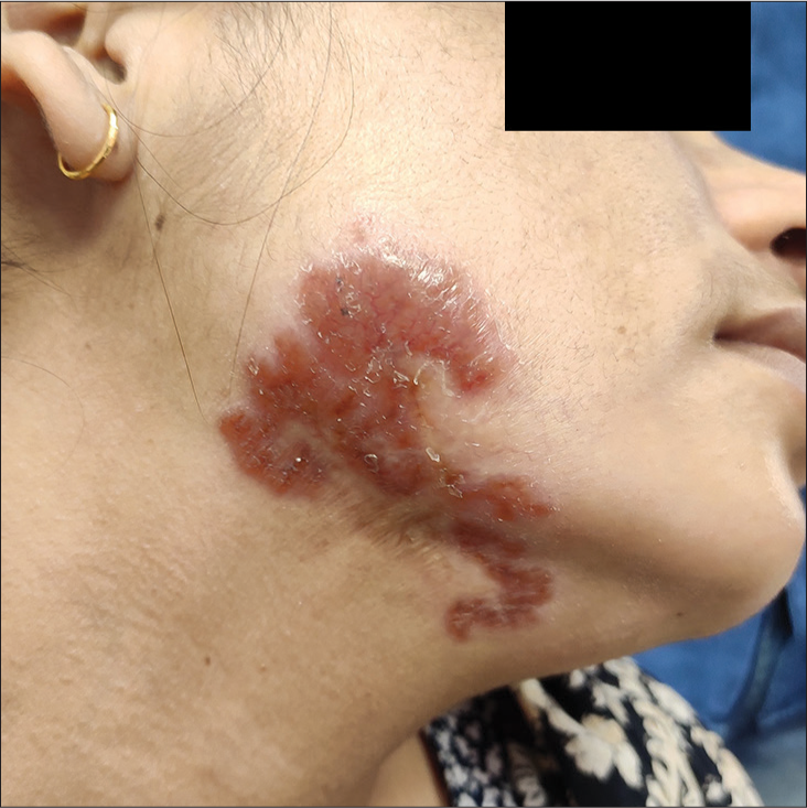
- Solitary, reddish-brown plaque presents over the lower right cheek, with scarring at the inferior margin and serpiginous progression at the superior margin.
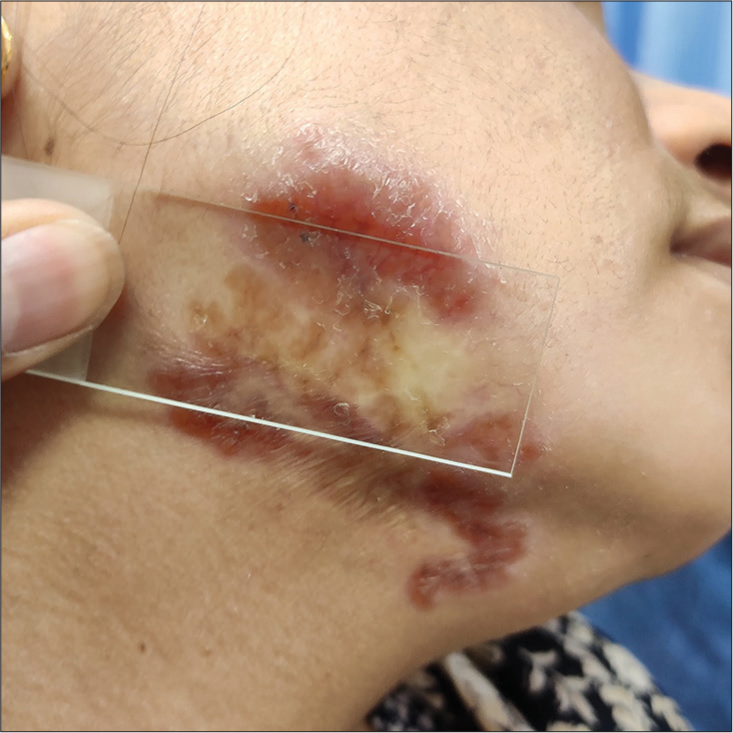
- Diascopy showing apple-jelly nodules.
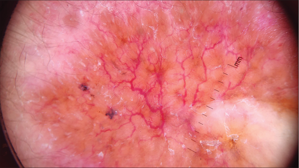
- Dermatoscopy showing linear focussed telangiectasias on a yellow-brown background and white scales (DermLite DL3N; dry, contact, polarised, ×10).
DIAGNOSIS AND DISCUSSION
Cutaneous tuberculosis (TB) constitutes around 1% of extrapulmonary TB. Lupus vulgaris (LV), also called as tuberculosis cutis luposa or boeck’s sarcoid, is a chronic, progressive, post-primary and paucibacillary form of cutaneous TB, occurring in a person with moderate to high immunity. It can arise either from an endogenous underlying focus or through exogenous inoculation. In India, buttocks and extremities are commonly affected, while in the West, facial involvement is commoner. It presents as brownish to erythematous, soft and solitary plaque, having an advancing edge and a scarring edge. It has several clinical variants such as papular, nodular, plaque, vegetative, ulcerative, atrophic, hypertrophic, mutilating and tumour-like.[1]
Lupoma is the characteristic dermatological lesion of LV. It can be well-demonstrated by diascopy or dermatoscopy, both of which show the ‘apple jelly’ or ‘suere d’orge’ (barley sugar) nodules.[2] Dermatoscopy, an intermediate between macroscopic and microscopic dermatology, reveals linear focussed telangiectasias on a yellow-brown background.[3] Yellow-brown areas are a hallmark of cutaneous granulomatous disorders, which include infectious granulomatous disorders, such as mycobacterial diseases, fungal diseases, leishmaniasis and syphilis, as well as non-infectious granulomatous disorders, such as sarcoidosis, necrobiotic disorders, interstitial granulomatous dermatitis, granulomatous rosacea and foreign body granulomas. The yellow-brown appearance is attributed to the mass effect of dermal granulomas. Apart from granulomatous disorders, similar yellow-brown areas have also been reported due to dense dermal cellular infiltration, such as xanthogranuloma, lymphomas and pseudo-lymphomas. The granulomas of LV are more yellowish than sarcoidosis because of the occasional presence of caseous necrosis, which makes the granuloma less compact, as well as due to the lipid deposition within the multinucleate Langhans giant cells.[3]
Jonathan Hutchinson first described the apple-jelly nodules. In 1865, he wrote, ‘Patches of lupus often present a very peculiar tint; they are at parts glossy and semi-transparent. Mr. Wilson (Erasmus Wilson) has well compared them to ‘reddish transparent jelly effused on the skin and streaked with the ramifications of a few small blood vessels.’[4] The term apple-jelly nodule was used to describe the ‘papules on plaque’ or the ‘tubers on plaque’ appearance of LV, with yellow-brown papules or nodules at the margins of the lesion, reminiscent of apple-jelly.[5] They have also been described as ‘apple-butter nodules.’[6] Linear or branching vessels seen on dermatoscopy signify the dilated dermal capillaries pushed up by the underlying granulomas. Other dermatoscopic features of LV include white structureless areas, white lines, follicular plugs, and scales.[7]
Ethical approval
Institutional Review Board approval is not required.
Declaration of patient consent
The authors certify that they have obtained all appropriate patient consent.
Conflicts of interest
There are no conflicts of interest.
Use of artificial intelligence (AI)-assisted technology for manuscript preparation
The authors confirm that there was no use of artificial intelligence (AI)-assisted technology for assisting in the writing or editing of the manuscript and no images were manipulated using AI.
Financial support and sponsorship
Nil.
References
- Lupus Vulgaris in a Mother and Child. BMJ Case Rep. 2021;14:e240591.
- [CrossRef] [PubMed] [Google Scholar]
- Dermatoscopy of Cutaneous Granulomatous Disorders. Indian Dermatol Online J. 2021;12:34-44.
- [CrossRef] [PubMed] [Google Scholar]
- The History of Lupus Vulgaris: Its Recognition, Nature, Treatment and Prevention In: Section of the History of Medicine. London: Proceedings of the Royal Society of Medicine; 1954. p. :127-8.
- [CrossRef] [Google Scholar]
- Apple-jelly Nodules of Cutaneous Rosai-Dorfman Disease. Indian Dermatol Online J. 2023;14:419-21.
- [CrossRef] [PubMed] [Google Scholar]
- Investigative and Clinical Studies with Diascopy in Dermatology. AMA Arch Dermatol. 1957;75:699-705.
- [CrossRef] [PubMed] [Google Scholar]
- Dermoscopy of the Diverse Spectrum of Cutaneous Tuberculosis in the Skin of Color. Dermatol Pract Concept. 2022;12:e2022203.
- [CrossRef] [PubMed] [Google Scholar]


