

Translate this page into:
Copper Penny Bodies in Chromoblastomycosis
*Corresponding author: Pankaj Das, Department of Dermatology, Armed Forces Medical College, Pune, Maharashtra, India. pankaj3609@gmail.com
-
Received: ,
Accepted: ,
How to cite this article: Das P, Vasudevan B, Singh GK, Sapra D. Copper Penny Bodies in Chromoblastomycosis. Indian J Postgrad Dermatol. 2024;2:48-9. doi: 10.25259/IJPGD_81_2023
A 63-year-old male presented with ulcerated lesions over the left leg of 3 years duration. Examination revealed verrucous plaques, erosions and ulcers covered with crusts with areas of scarring [Figure 1]. A 10% potassium hydroxide mount revealed small round light-brown coloured bodies lying singly and in clusters called copper penny bodies, medlar bodies and muriform or sclerotic cells diagnostic of chromoblastomycosis [Figure 2]. Chromoblastomycosis is a subcutaneous mycosis caused by a group of dematiaceous (pigment-producing) fungi.[1] Medlar bodies are thick-walled cells (5–12 microns) with multiple internal transverse septa or chambers that resemble copper pennies.[2]

- Left leg showing verrucous plaques, erosions, ulcers covered with crusts with areas of scarring, and post-inflammatory hyperpigmentation.
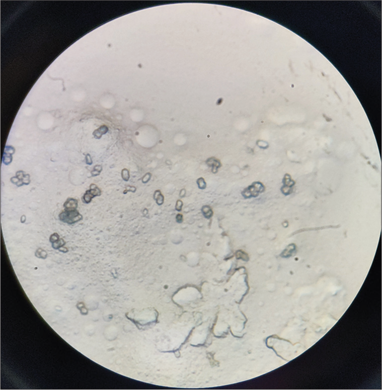
- 10% potassium hydroxide (KOH) mount showing small round light brown coloured bodies lying singly and in groups called copper penny bodies, medlar bodies and muriform or sclerotic cells diagnostic of chromoblastomycosis (×100, KOH).
Ethical approval
The research was in compliance with Helsinki declaration 1964.
Declaration of patient consent
Patient’s consent not required as patients identity is not disclosed or compromised.
Conflicts of interest
There are no conflicts of interest.
Use of artificial intelligence (AI)-assisted technology for manuscript preparation
The authors confirm that there was no use of artificial intelligence (AI)-assisted technology for assisting in the writing or editing of the manuscript and no images were manipulated using AI.
Financial support and sponsorship
Nil.
References
- Chromoblastomycosis: An Etiological, Epidemiological, Clinical, Diagnostic, and Treatment Update. An Bras Dermatol. 2018;93:495-506.
- [CrossRef] [PubMed] [Google Scholar]
- Cytodiagnostic Copper Pennies in Chromoblastomycosis. Indian Dermatol Online J. 2016;7:145-46.
- [CrossRef] [PubMed] [Google Scholar]


