Translate this page into:
Nail-Fold Capillaroscopy Quiz
*Corresponding author: Vishal Gaurav, Department of Dermatology and Venereology, Maulana Azad Medical College, New Delhi, India. mevishalgaurav@gmail.com
-
Received: ,
Accepted: ,
How to cite this article: Gaurav V. Nail-Fold Capillaroscopy Quiz. Indian J Postgrad Dermatol. doi: 10.25259/IJPGD_92_2024
QUESTIONS
-
Which of the following is NOT a feature of the ‘Early’ nail-fold videocapillaroscopy (NVC) pattern in systemic sclerosis?
Few dilated and/or giant capillaries
Avascular areas
Few haemorrhages
Preserved distribution
-
The presence of bushy and ramified capillaries is characteristic of which NVC pattern of systemic sclerosis?
Early NVC Pattern
Active NVC Pattern
Late NVC Pattern
Both A and B
-
According to the LeRoy and Medsger criteria, which of the following is required for diagnosing primary Raynaud’s phenomenon?
Asymmetrical involvement
Presence of tissue necrosis
Normal capillaroscopic pattern
Positive test for antinuclear antibodies
-
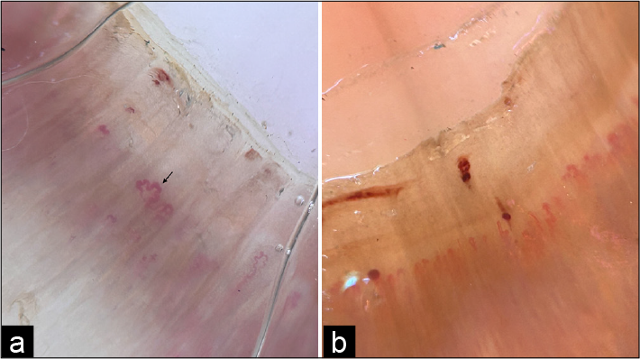
Which of the following best describes the capillaries shown in Figure 1a, marked by the black arrow?
Dilated capillaries
Tortuous capillaries
Meandering capillaries
Both (a) and (b)
 Figure 1:
- Nailfold capillaroscopy (Dermlite DL5) of (a) Left middle fingernail, capillary marked by a black arrow (polarised; ×20); (b) Middle fingernail (polarised; ×20).
-
Which nailfold capillaroscopy (NFC) pattern is likely to be observed in healthy individuals?
Meandering capillaries
Bushy capillaries
Dilated subpapillary plexus
Increased tortuosity (>10%)
-
Which NVC pattern is shown in Figure 1b?
Early NVC Pattern
Active NVC Pattern
Late NVC Pattern
Intermediate Pattern
-
What is a normal variant observed in NFC?
Increased tortuosity in 10% of capillaries
Criss-cross forms
Presence of haemorrhages
All of the above
-
What is the recommended avoidance time for caffeine and smoking before performing NFC?
2–4 h
4–6 h
6–8 h
8–10 h
-
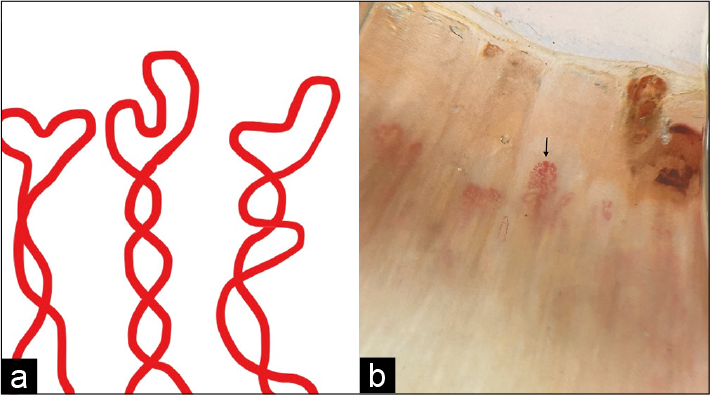
Which of the following best describes the capillaries shown in schematic Figure 2a?
Dilated capillaries
Tortuous capillaries
Meandering capillaries
Bushy capillaries
 Figure 2:
- (a) Schematic representation of microvascular architectural abnormality; (b) Nail fold capillaroscopy (Dermlite DL5) of left ring fingernail, capillary marked by a black arrow (polarised; ×20).
-
What is the diagnostic significance of devascularisation and architectural distortion on NVC in systemic sclerosis?
Early diagnosis
Indicator of disease activity
Occurrence of digital ulcers
Both a and b
-
Which of the following statements is true regarding capillary density in NFC?
Higher density in toes than fingers
Decreases with age
Influenced by gender
Difficult to calculate in darker skin
-
Which observation is typical for NFC in systemic lupus erythematosus (SLE)?
Microhaemorrhages
Elongated and meandering capillaries
Bushy capillaries
Presence of avascular areas
-
What characterises dystrophic capillary loops in NFC?
Normal size and caliber
Abnormally slow blood flow
Malformed and not well developed
Increased capillary density
-
Which parameter is defined as the maximum open space measured at the apex of a capillary?
Capillary width
Apex width
Internal diameter
Arterial limb diameter
-
Which capillary morphology is shown in Figure 2b, marked by the black arrow?
Meandering capillaries
Bushy capillaries
Bizarre capillaries
Dystrophic capillaries
-
Which finding is associated with NFC in patients with dermatomyositis?
Elongated capillaries
Dilated capillaries and tortuous vessels
Increased density of capillaries
Both a and b
-
Which of the following is true about the correlation between capillary abnormalities and organ involvement in systemic sclerosis?
No correlation exists
Correlation exists with skin involvement only
Correlation exists with both cutaneous and visceral involvement
Correlation exists with disease duration only
-
Which statement is correct regarding NFC in patients with antiphospholipid syndrome (APS)?
NFC shows normal capillary patterns
NFC reveals increased capillary density
NFC may show capillary haemorrhages
NFC is not useful in APS
-
What is the gold standard technique for morphological assessment of nail-fold capillaries?
Video capillaroscope
Stereomicroscope
Wide-field microscope
Universal serial bus (USB) dermatoscope
-
NFC is useful in which of the following paediatric diseases?
Kindler’s syndrome
Fabry’s disease
Kawasaki disease
All of the above
ANSWERS
-
b
In systemic sclerosis, the ‘Early’ NVC pattern is characterised by the presence of a few dilated and/or giant capillaries, few haemorrhages and preserved capillary distribution. Avascular areas are typically absent in the early stage and appear in the ‘Active’ or ‘Late’ NVC patterns, reflecting more advanced microvascular damage.[1,2]
-
c
Bushy and ramified capillaries are a hallmark of the ‘Late’ NVC pattern in systemic sclerosis. They indicate advanced microangiopathy, reflecting significant neoangiogenesis and capillary remodelling. This feature is not seen in the ‘Early’ or ‘Active’ patterns, which are characterised by less severe vascular changes, such as dilated capillaries and haemorrhages in the early stages or capillary loss and avascular areas in the active stage.[1,2]
-
c
According to the LeRoy and Medsger criteria, a normal capillaroscopic pattern is required for diagnosing primary Raynaud’s phenomenon, as it distinguishes it from secondary forms associated with underlying connective tissue diseases. Secondary Raynaud’s often shows abnormal capillaroscopy findings, such as giant capillaries or avascular areas. The other options – asymmetrical involvement, tissue necrosis and positive anti-nuclear antibody (ANA) – are indicative of secondary Raynaud’s phenomenon.[3,4]
-
d
The capillaries in Figure 1a exhibit features of both dilation (widened loops) and tortuosity (twisting or bending of capillaries), which are commonly seen together in systemic sclerosis. Dilated capillaries appear as enlarged loops, while tortuous capillaries display irregular, winding shapes. Both features can coexist and are often described together in NFC findings.[2]
-
c
In healthy individuals, NFC typically shows a normal capillary morphology, and slight dilation of the subpapillary plexus is a common finding, seen in 26% of healthy individuals. This is a physiological variation and does not indicate pathology. In contrast, features such as meandering capillaries, bushy capillaries and increased tortuosity >10% are associated with microvascular abnormalities or underlying diseases.[5]
-
b
The active NVC pattern in systemic sclerosis is characterised by features such as the presence of giant capillaries, frequent haemorrhages, moderate capillary loss and the beginning of architectural disorganisation [Figure 1b]. These findings distinguish it from the Early pattern (minimal changes) and the Late pattern (severe capillary loss and bushy capillaries).[1,2]
-
b
Criss-cross forms are a recognised normal variant in NFC and do not indicate pathology. Criss-cross forms, or figure-of-eight capillaries, are characterised by crossed arterial and venous limbs. In contrast, features such as increased tortuosity in 10% of capillaries and presence of haemorrhages are typically associated with microvascular abnormalities or underlying conditions and are not considered normal.[6]
-
b
It is recommended to avoid caffeine and smoking for 4–6 h before performing NFC because both substances can cause transient microvascular changes, such as vasoconstriction or altered capillary flow, which may interfere with accurate assessment of the capillaries. This time frame ensures the vascular system returns to its baseline state for reliable results.[2]
-
c
Meandering capillaries are described as capillaries with limbs that cross over themselves or other capillaries multiple times [Figure 2a]. This distinct morphology differentiates them from tortuous capillaries, which are curled without crossing over themselves, and bushy capillaries, which show multiple buds or branches originating from a single capillary loop. Bizarre capillaries are atypical and do not fit into defined categories.[2,6,7]
-
c
Devascularisation and architectural distortion on NVC in systemic sclerosis indicate advanced microvascular damage. These changes are strongly associated with the occurrence of digital ulcers, a common and severe complication in systemic sclerosis. While early NVC patterns help in early diagnosis and identifying disease activity, extensive capillary loss and disorganisation are more predictive of ischaemic complications like ulcers.[8]
-
d
Capillary density assessment in NFC can be challenging in individuals with darker skin due to increased pigmentation, which may obscure visibility of the capillaries. This contrasts with other options: Capillary density is higher in fingers than toes, does not significantly decrease with age and is not influenced by gender. Thus, pigmentation-related difficulty is the most accurate statement.[9]
-
b
In SLE, NFC typically reveals elongated and meandering capillaries due to microvascular changes associated with the disease. These findings are characteristic of SLE and differ from other connective tissue diseases like systemic sclerosis, where findings such as bushy capillaries or avascular areas are more common. While microhaemorrhages may occur, they are not specific to SLE.[10]
-
c
Dystrophic capillary loops are characterised by their malformation and underdevelopment, reflecting structural damage to the microvasculature. They appear irregular, poorly formed and sometimes fragmented. This distinguishes them from normal capillaries, which have consistent size and caliber, and from capillaries with abnormally slow blood flow, which may indicate functional rather than structural issues. Increased capillary density is not associated with dystrophic changes but rather with neoangiogenesis or other processes.[11,12]
-
b
The apex width refers to the maximum open space measured at the apex of a capillary loop, representing the widest point of the capillary’s uppermost curve. This parameter is specifically used in NFC to assess capillary dilation. It is distinct from capillary width, which considers the overall dimensions, and internal diameter or arterial limb diameter, which measure other aspects of capillary structure.[2]
-
b
Bushy capillaries are characterised by multiple small buds or branches originating from a single capillary loop, giving them a bushy or ramified appearance [Figure 2b]. This morphology occurs due to neoangiogenesis. Meandering capillaries have loops that cross over themselves or others. Bizarre capillaries exhibit atypical and irregular shapes, while dystrophic capillaries are malformed and poorly developed.[2,6,7]
-
d
In dermatomyositis, NFC typically shows elongated capillaries, which are often seen as abnormal extensions of the capillary loops, and dilated capillaries and tortuous vessels, which indicate microvascular damage and dysfunction. These findings are characteristic of the capillary abnormalities seen in dermatomyositis, reflecting the underlying vascular involvement in the disease. Increased density of capillaries is not a typical finding in dermatomyositis.[10]
-
c
In systemic sclerosis, capillary abnormalities observed in NFC are strongly correlated with both cutaneous and visceral involvement. These microvascular changes, such as dilated or avascular capillaries, can reflect the severity and extent of skin fibrosis as well as internal organ involvement, particularly the lungs, heart and gastrointestinal tract. The capillary pattern provides insights into the overall disease severity and can help in predicting the risk of complications, including organ damage.[13,14]
-
c
In patients with APS, NFC can reveal capillary haemorrhages, which are small areas of blood leakage from capillaries. This finding is indicative of microvascular damage, a common complication in APS due to hypercoagulability and vascular thrombosis. While increased capillary density is more typical of other conditions such as systemic sclerosis, normal capillary patterns and NFC being not useful are incorrect because NFC can provide valuable information about vascular changes in APS.[15]
-
a
The video capillaroscope is considered the gold standard technique for the morphological assessment of nail-fold capillaries. It provides high-resolution, real-time imaging, allowing detailed visualisation of capillary loops and abnormalities in microcirculation. This technique is preferred because it allows for precise measurement and documentation of capillary morphology, such as dilation, tortuosity and avascular areas. Other methods such as the stereomicroscope, wide-field microscope and USB dermatoscope are also used but do not offer the same level of detailed morphological analysis as video capillaroscopy.[2]
-
d
NFC is a valuable diagnostic tool for assessing microvascular changes in paediatric diseases, including Kindler’s syndrome, Fabry’s disease and Kawasaki disease. In Kindler’s syndrome, NFC may show reduced capillary density, neoangiogenesis and the presence of dilated or giant capillaries. Fabry’s disease can present with tortuous and dilated capillaries due to systemic vascular involvement. In Kawasaki disease, NFC reveals reduced capillary density, an increase in limb diameter and greater inter-capillary distance, reflecting vascular changes linked to inflammation.[2,16,17]
Ethical approval
Institutional Review Board approval is not required.
Declaration of patient consent
The authors certify that they have obtained all appropriate patient consent.
Conflicts of interest
There are no conflicts of interest.
Use of artificial intelligence (AI)-assisted technology for manuscript preparation
The author confirms that there was no use of artificial intelligence (AI)-assisted technology for assisting in the writing or editing of the manuscript and no images were manipulated using AI.
Financial support and sponsorship: Nil.
References
- Nailfoldvideocapillaroscopy Assessment of Microvascular Damage in Systemic Sclerosis. J Rheumatol. 2000;27:155-60.
- [Google Scholar]
- Nail-fold Capillaroscopy for the Dermatologists. Indian J Dermatol Venereol Leprol. 2022;88:300-12.
- [CrossRef] [PubMed] [Google Scholar]
- Raynaud's Phenomenon: A Proposal for Classification. Clin Exp Rheumatol. 1992;10:485-8.
- [Google Scholar]
- Nailfold Digital Capillaroscopy in 447 Patients with Connective Tissue Disease and Raynaud's Disease. J Eur Acad Dermatol Venerol. 2004;18:62-8.
- [CrossRef] [PubMed] [Google Scholar]
- Quantitative Nailfold Capillaroscopy Findings in a Population with Connective Tissue Disease and in Normal Healthy Controls. Ann Rheum Dis. 1996;55:507-12.
- [CrossRef] [PubMed] [Google Scholar]
- Capillaroscopic Observations in Rheumatic Diseases. Ann Rheum Dis. 1970;29:244-53.
- [CrossRef] [PubMed] [Google Scholar]
- The Contribution of Capillaroscopy to the Differential Diagnosis of Connective Autoimmune Diseases. Best Pract Res Clin Rheumatol. 2007;21:1093-108.
- [CrossRef] [PubMed] [Google Scholar]
- Position Article and Guidelines 2018 Recommendations of the Brazilian Society of Rheumatology for the Indication, Interpretation and Performance of Nailfold Capillaroscopy. Adv Rheumatol. 2019;59:5.
- [CrossRef] [PubMed] [Google Scholar]
- How to Perform and Interpret Capillaroscopy. Best Pract Res Clin Rheumatol. 2013;27:237-48.
- [CrossRef] [PubMed] [Google Scholar]
- The Handheld Dermatoscope as a Nail-fold Capillaroscopic Instrument. Arch Dermatol. 2003;139:1027-30.
- [CrossRef] [PubMed] [Google Scholar]
- Nail Fold Capillaroscopy in the Study of Microcirculation in Elderly Hypertensive Patients. Arch Gerontol Geriatr. 1996;5:79-83.
- [CrossRef] [PubMed] [Google Scholar]
- Clinical Applicability of Quantitative Nailfold Capillaroscopy in Differential Diagnosis of Connective Tissue Diseases with Raynaud's Phenomenon. J Formos Med Assoc. 2013;112:482-8.
- [CrossRef] [PubMed] [Google Scholar]
- Nailfold Capillaroscopy is Useful for the Diagnosis and Follow-up of Autoimmune Rheumatic Diseases. A Future Tool for the Analysis of Microvascular Heart Involvement? Rheumatology (Oxford) 2006. ;. ;45:43-6.
- [CrossRef] [PubMed] [Google Scholar]
- The Complexity of Managing Systemic Sclerosis: Screening and Diagnosis. Rheumatology (Oxford). 2009;48(Suppl 3):iii8-13.
- [CrossRef] [PubMed] [Google Scholar]
- Nailfold Videocapillaroscopy in Primary Antiphospholipid Syndrome (PAPS) Rheumatology. 2004;43:1025-7.
- [CrossRef] [PubMed] [Google Scholar]
- Nailfold Capillaroscopic Changes in Kindler Syndrome. Intractable Rare Dis Res. 2015;4:214-6.
- [CrossRef] [PubMed] [Google Scholar]
- Deterioration of Cutaneous Microcirculatory Status of Kawasaki Disease. Clin Rheumatol. 2012;31:847-52.
- [CrossRef] [PubMed] [Google Scholar]


