Charcot’s Arthropathy in Leprosy
*Corresponding author: Pankaj Das, Department of Dermatology, Armed Forces Medical College, Pune, Maharashtra, India. pankaj3609@gmail.com
-
Received: ,
Accepted: ,
How to cite this article: Das P, Bhatnagar A, Sharma NK. Charcot’s Arthropathy in Leprosy. Indian J Postgrad Dermatol. doi: 10.25259/IJPGD_3_2025
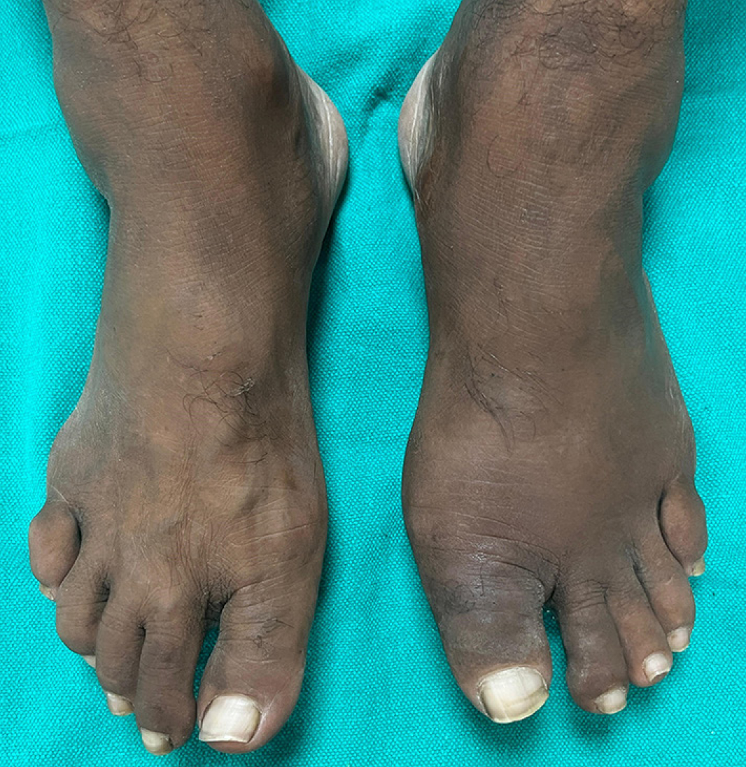
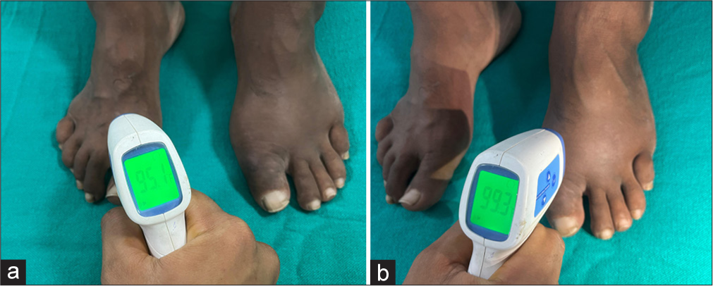
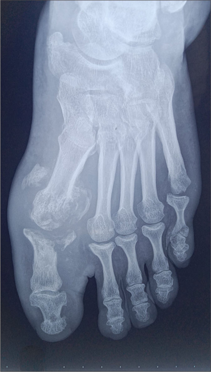
A 46-year-old male, a known case of lepromatous leprosy on regular multi-drug therapy for the past 6 months presented with painless swelling of the left foot for 3 weeks duration. The patient provided a history of numb legs with painless recurrent trophic ulcers on both feet for the past 2 years. Clinical examination revealed a mild but diffuse swelling affecting the left forefoot [Figure 1]. The left forefoot was warm to touch, and on measuring temperature with an infra-red thermometer, it was 4.2°F warmer than the contralateral side (99.3°F vs. 95.1°F) [Figure 2]. X-ray of the left foot revealed erosion of the first metatarsophalangeal joint with multiple intra-articular loose bodies with distension of joint space and juxta-articular osteopenia. Similar changes were also noted in the fifth metatarsophalangeal joint [Figure 3]. The clinical signs corroborated by radiological findings are classical to Charcot’s arthropathy. A red-hot swollen foot in the absence of symptoms is characteristic of Charcot arthropathy. The stages progress through prodrome, destruction, coalescence and consolidation. Our patient was in the joint destruction stage, which was confirmed by classic findings in the X-ray. Treatment is mainly non-surgical by promptly immobilising the foot and offloading by application of total contact plaster cast to restrict weight-bearing aiming at preventing permanent deformity till the signs of inflammation subsides which usually take 6–8 weeks. Bisphosphonates are indicated in the acute phase as they inhibit osteoclastic reabsorption.

- Diffuse swelling of the left foot and the first two toes. Erythema is not appreciable in skin of colour.

- (a and b) Measurement of temperature with an infra-red thermometer- right foot 95.1°F, left foot 99.3°F (4.2°F warmer than the contralateral side).

- Dorsoplantar X-ray of the left foot revealed erosion of first metatarsophalangeal joint with multiple intraarticular loose bodies with distention of joint space and juxta-articular osteopenia. Similar changes were also noted in the fifth metatarso-phalangeal joint.
Ethical approval
Institutional Review Board approval is not required.
Declaration of patient consent
The authors certify that they have obtained all appropriate patient consent.
Conflicts of interest
There are no conflicts of interest.
Use of artificial intelligence (AI)-assisted technology for manuscript preparation
The authors confirm that there was no use of artificial intelligence (AI)-assisted technology for assisting in the writing or editing of the manuscript and no images were manipulated using AI.
Financial support and sponsorship: Nil.


