Translate this page into:
Trich KY Nodule on the Nose
*Corresponding author: Sanjanaa Srinivasa, Department of Dermatology, St John’s Medical College, Bengaluru, Karnataka, India. sanjanaa.srinivasa@stjohns.in
-
Received: ,
Accepted: ,
How to cite this article: Srinivasa S, Lobo C, Kaimal S, Malipatel R. Trich KY Nodule on the Nose. Indian J Postgrad Dermatol. 2025;3:62-3. doi: 10.25259/IJPGD_189_2024
CASE DESCRIPTION
A 52-year-old female presented with a skin coloured, minimally painful swelling over the nose, gradually increasing in size over the past 5 years. On inquiry, the patient did not have any significant medical or surgical history in the past.
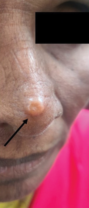
On examination, a skin coloured firm, non-tender nodule measuring 1 × 1 cm was noted on the dorsum of the nose [Figure 1]. Differential diagnosis considered was trichoblastoma, basal cell carcinoma and nodular amyloidosis.

- Solitary, skin coloured nodule on the nose (black arrow).
HISTOPATHOLOGIC FINDINGS
An excision biopsy was done and histopathology revealed multilobulated, circumscribed neoplasm composed of bland trichoblastic cells arranged in nests, cords, reticular and cribriform pattern, with no evidence of mitosis or atypia [Figure 2].
![(a) Dermis shows a multilobulated, circumscribed neoplasm arranged as nests, cords, reticular and cribriform pattern, with few papillary mesenchymal bodies (haematoxylin and eosin [H & E] ×2). (b) Bland appearing trichoblastic cells with peripheral palisading, with no evidence of atypia or mitosis (H & E ×20).](/content/146/2025/3/1/img/IJPGD-3-062-g002.png)
- (a) Dermis shows a multilobulated, circumscribed neoplasm arranged as nests, cords, reticular and cribriform pattern, with few papillary mesenchymal bodies (haematoxylin and eosin [H & E] ×2). (b) Bland appearing trichoblastic cells with peripheral palisading, with no evidence of atypia or mitosis (H & E ×20).
DIAGNOSIS
Trichoblastoma.
DISCUSSION
Trichoblastoma is an uncommon benign, biphasic adnexal neoplasm originating from follicular germ cells. It presents as an asymptomatic, solitary, well-circumscribed, skin-coloured papule or nodule, most commonly in elderly individuals. It usually occurs over the head-and-neck region, trunk and extremities.[1]
Histopathological variants include nodular, racemiform, columnar, retiform, cribriform, clear cell, pigmented and adamantinoid patterns.[2]
The closest differential diagnosis clinically and dermoscopically includes basal cell carcinoma (BCC). It is imperative to distinguish between both as trichoblastoma is typically benign and BCC exhibits malignant behaviour.
BCC originates from basal layer of epidermis. It has a polymorphic clinical presentation, which includes nodular superficial, morphoeic (sclerosing), keratotic, cystic, pigmented and micronodular variants.[3] Histopathological features include prominent peripheral palisading, artefactual clefting, increased mitotic figures, variable necrosis, calcification, myxoinflammatory stroma, lymphocytic infiltrate and intraepithelial cluster of differentiation (CD) 10 staining.[1,4]
Trichoblastoma exhibits peripheral palisading, keratin cysts, no artefactual clefting, absent or focal cellular atypia and necrosis, variable stromal condensation, follicular papillae and peritumoural stromal CD10 staining.[1,4]
On dermoscopy, small fine arborizing vessels and spoke-wheel areas are present in trichoblastoma more frequently than BCC. In BCC, wider branching telangiectasia, rainbow pattern, blue-grey ovoid nests and blue-grey globules are more common.[4]
Dermoscopic differences are not exclusive for either trichoblastoma or BCC; hence, histology remains the gold standard for diagnosis.
Trichoblastoma is usually benign with rare malignant transformation. Surgical excision is the preferred therapeutic modality.[5]
Ethical approval
Institutional Review Board approval is not required.
Declaration of patient consent
The authors certify that they have obtained all appropriate patient consent.
Conflicts of interest
There are no conflicts of interest.
Use of artificial intelligence (AI)-assisted technology for manuscript preparation
The authors confirm that there was no use of artificial intelligence (AI)-assisted technology for assisting in the writing or editing of the manuscript and no images were manipulated using AI.
Financial support and sponsorship
Nil.
References
- Skin Adnexal Neoplasms-Part 1: An Approach to Tumours of the Pilosebaceous Unit. J Clin Pathol. 2007;60:129-44.
- [CrossRef] [PubMed] [Google Scholar]
- The Multiple Faces of Nodular Trichoblastoma: Review of the Literature with Case Presentation. Dermatopathology (Basel). 2021;8:265-70.
- [CrossRef] [PubMed] [Google Scholar]
- Guidelines for the Management of Basal Cell Carcinoma. Br J Dermatol. 2008;159:35-48.
- [CrossRef] [PubMed] [Google Scholar]
- Trichoblastoma: Is a Clinical or Dermoscopic Diagnosis Possible? J Eur Acad Dermatol Venereol. 2016;30:1978-80.
- [CrossRef] [PubMed] [Google Scholar]


