Translate this page into:
Oral Mucous Patch of Secondary Syphilis
*Corresponding author: Rajendra Devanda, Department of Dermatology Venereology and Leprosy, National Institute of Medical Sciences and Research, Jaipur, Rajasthan, India. rdevanda24@gmail.com
-
Received: ,
Accepted: ,
How to cite this article: Couppoussamy K, Devanda R. Oral Mucous Patch of Secondary Syphilis. Indian J Postgrad Dermatol. 2025;3:74-5. doi: 10.25259/IJPGD_163_2024
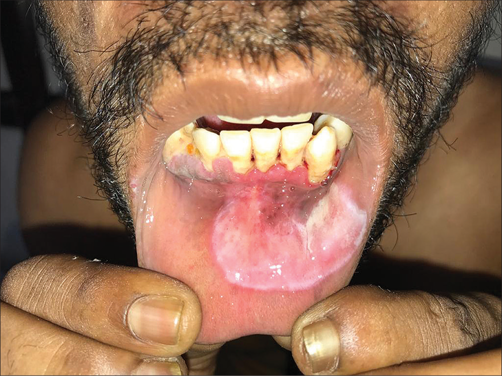
A 35-year-old male presented with an asymptomatic white lesion in the oral cavity for 2 weeks. The patient had unprotected sexual intercourse with an unknown female partner 3 months ago. There was no history of genital lesions in the past. On examination, single large 3 × 3 cm2 whitish mucosal patch was present over the lower lip with erythema of gums and bleeding points [Figure 1]. No other skin or genital lesions were noted in the patient. A venereal disease research laboratory test was positive with 1:32 dilution, and the Treponema pallidum haemagglutination assay test was positive. Other investigations such as human immunodeficiency virus, hepatitis C virus and hepatitis B surface antigen were non-reactive. Thus, a diagnosis of secondary syphilis was made. The oral lesion is a mucous patch seen in secondary syphilis. The patient was treated with a benzathine penicillin 2.4 million units as single intramuscular dose. At the 1-month follow-up, the lesion healed completely. A higher degree of suspicion is needed to diagnose such cases when the presentation is only an oral lesion without any other skin lesions. Syphilis is caused by T. pallidum with varied kind of clinical manifestations. They can present with cutaneous or mucosal lesions. The lesions in the oral cavity of secondary syphilis includes ulcers such as snail track, mucous patch and condyloma lata presenting as split papules at the angles of mouth.

- Single large 3 × 3 cm whitish mucosal patch noted over the lower lip with erythema of the gum and bleeding spots.
Ethical approval
Institutional Review Board approval is not required.
Declaration of patient consent
The authors certify that they have obtained all appropriate patient consent.
Conflicts of interest
There are no conflicts of interest.
Use of artificial intelligence (AI)-assisted technology for manuscript preparation
The authors confirm that there was no use of artificial intelligence (AI)-assisted technology for assisting in the writing or editing of the manuscript and no images were manipulated using AI.
Financial support and sponsorship
Nil.


