

Translate this page into:
Moniliform Blepharosis in Lipoid Proteinosis
*Corresponding author: Pankaj Das, Department of Dermatology, Armed Forces Medical College, Pune, Maharashtra India. pankaj3609@gmail.com
-
Received: ,
Accepted: ,
How to cite this article: Das P, Bhatnagar A, Krishnan L, Kunwar B, Choudhary SR. Moniliform Blepharosis in Lipoid Proteinosis. Indian J Postgrad Dermatol. doi: 10.25259/IJPGD_65_2025
A 22-year-old male, born of non-consanguineous marriage presented to us with hoarseness of voice with multiple papules over both eyelids since childhood. The eyelid lesions began as skin coloured tiny asymptomatic swellings, progressing to a beading all along the margins of the eye lashes. Dermatological examination revealed multiple, skin coloured, linearly arranged, closely aggregated 1–2 mm sized papules predominantly along the eyelid margins (moniliform blepharosis) [Figure 1a]. There were round to linear atrophic scars over the face. Oral examination showed a coarse, thickened and scarred mucosa of the palate, uvula and buccal mucosa [Figure 1b]. Genetic analysis confirmed a homozygous non-sense mutation in exon 3 of the extracellular matrix protein 1 (ECM1) gene (c.157 C>T). Based on the clinical and genetic testing, the patient was diagnosed as a case of lipoid proteinosis (LP). He was started on tab acitretin at a dose of 25 mg/day. Three months after the initiation of therapy, there was some regression and softening of lesions on the eyelids but no improvement in the hoarseness of voice.
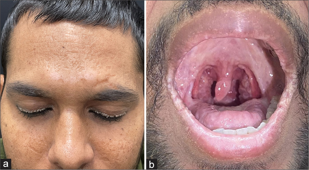
- (a) There is involvement of eyelids in form of multiple, skin coloured, linearly arranged, closely aggregated 1–2 mm sized papules (moniliform blepharosis). The face also shows atrophic scars. (b) Oral examination shows a coarse, thickened and scarred mucosa of the palate and uvula.
Lipoid proteinosis (LP) is a rare autosomal recessive genodermatosis characterized by deposition of Periodic Acid-Schiff (PAS)-positive hyaline material in skin, mucosa, and other tissues.[1] It is caused by loss-of-function mutation in the ECM1 gene coding extracellular matrix protein 1.[2] The eyelids and mucosae are mainly affected with beading and scarring respectively. The mucosal disease also affects the vocal cords which causes hoarseness of voice at an early age. Moniliform blepharosis is pathognomic of LP.
Ethical approval:
Institutional Review Board approval is not required.
Declaration of patient consent:
The authors certify that they have obtained all appropriate patient consent.
Conflicts of interest:
There are no conflicts of interest.
Use of artificial intelligence (AI)-assisted technology for manuscript preparation:
The authors confirm that there was no use of artificial intelligence (AI)-assisted technology for assisting in the writing or editing of the manuscript and no images were manipulated using AI.
Financial support and sponsorship: Nil.


