

Translate this page into:
Bedside Demonstration of Fluorescence Using 365 nm Wood`s Lamp Mode of Dermoscope in Pityriasis Versicolor
*Corresponding author: Shrinivas Patil, Department of Dermatology, Venereology and Leprosy, Command Hospital Air Force Bengaluru, Bengaluru, Karnataka, India. shrinivas93doctor@gmail.com
-
Received: ,
Accepted: ,
How to cite this article: Patil S, Bhatnagar A, Mitra D. Bedside Demonstration of Fluorescence Using 365 nm Wood`s Lamp Mode of Dermoscope in Pityriasis Versicolor. Indian J Postgrad Dermatol. 2024;2:150-1. doi: 10.25259/IJPGD_46_2024
Pityriasis versicolor, although clinical diagnosis is usually made by the presence of hypo or hyperpigmented scaly macules [Figure 1], can sometimes present with atypical features, necessitating additional diagnostic modalities. Wood’s lamp examination, particularly in the fluorescence can aid in the rapid diagnosis of this condition by highlighting the characteristic yellow-green fluorescence of the causative organism, Malassezia spp.
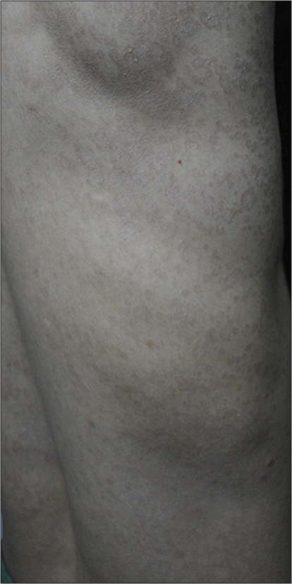
- Multiple discrete to coalescing hyperpigmented brownish scaly macules distributed over the right side of trunk.
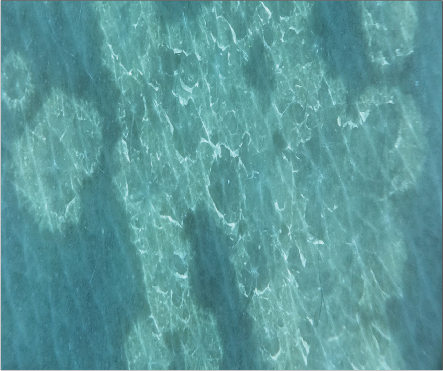
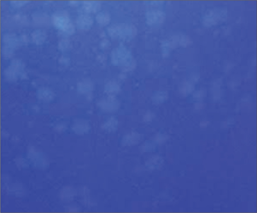
Dermoscopy, with its ability to magnify and visualise subtle skin changes, further enhances the diagnostic accuracy of Wood’s lamp examination, especially at the bedside. Bedside demonstration of fluorescence using the 365 nm Wood’s lamp mode of dermoscopy [Figure 2] is a valuable tool in the diagnosis of pityriasis versicolor, offering a rapid and non-invasive means of confirming the clinical suspicion. The green-yellow fluorescence observed in pityriasis versicolor lesions under Wood’s light is attributed to the presence of pityrialactone [Figure 3]. Figure 4 shows photograph of pityriasis versicolor as seen through woods lamp magnifier showing green-yellow fluorescence. Early diagnosis facilitated by this technique allows for the timely initiation of appropriate therapy, thereby improving patient outcomes and reducing the risk of complications.

- Demonstration of fluorescence using non-contact dermoscopy (DERMLITE DL5, 365 nm Woods lamp mode) showing yellow-green fluorescence.
- Contact dermoscopy (DERMLITE DL5, 365 nm woods lamp mode, ×10) showing yellow-green fluorescence.
- Photograph showing green-yellow fluorescence as seen through woods lamp magnifier.
Ethical approval
Institutional Review Board approval is not required.
Declaration of patient consent
The authors certify that they have obtained all appropriate patient consent.
Conflicts of interest
There are no conflicts of interest.
Use of artificial intelligence (AI)-assisted technology for manuscript preparation
The authors confirm that there was no use of artificial intelligence (AI)-assisted technology for assisting in the writing or editing of the manuscript and no images were manipulated using AI.
Financial support and sponsorship
Nil.


